Research
Most of my research focuses on MRI acquisition and post-processing methods for studying tissue microstructure in the brain. My current goals are to measure microstructure with high precision, to understand the cellular and molecular basis of these measurements by comparing them with histology, and to use them in patient studies to learn how brain diseases develop.
Physics-informed deep learning
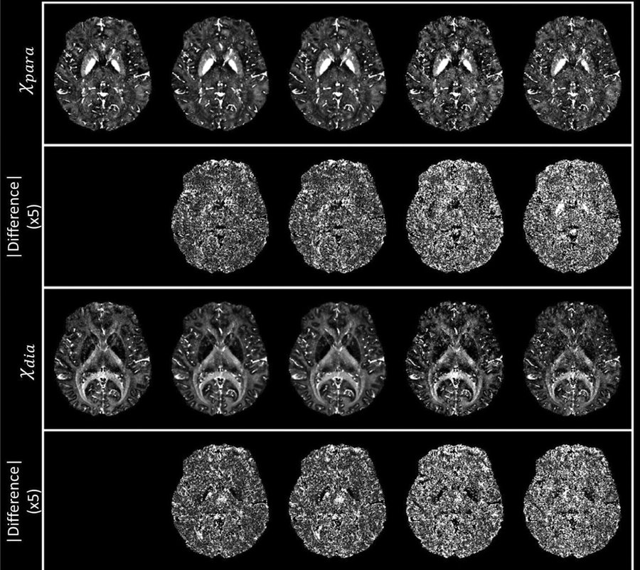
Advanced quantitative maps often need long scans or heavy computation, which limits their use in the clinic. I develop neural networks that are trained with physics-based models, so they produce accurate maps from shorter protocols and stay reliable on data that differ from the training set.
Examples include χ-sepnet, which produces χ-separation maps from multi-echo gradient-echo data alone, QSM dipole inversion networks that generalize across image resolutions, wavelet-based and proximal multi-operator networks for susceptibility imaging, and DeepResp for correcting respiration-induced artifacts.
Related papers
-
χ-sepnet: deep neural network for magnetic susceptibility source separation
Human Brain Mapping 46(2):e70136 (2025) *Co-corresponding authors
-
Resolution generalization of deep learning-based dipole inversion networks for QSM
NeuroImage 324:121598 (2025) *Corresponding authors
-
WaveSep: a flexible wavelet-based approach for source separation in susceptibility imaging
Machine Learning in Clinical Neuroimaging, Lecture Notes in Computer Science 14312:56-66 (2023) †Co-first authors
-
DeepResp: deep learning solution for respiration-induced B0 fluctuation artifacts in multi-slice GRE
NeuroImage 224:117432 (2021)
Iron and myelin imaging (χ-separation)
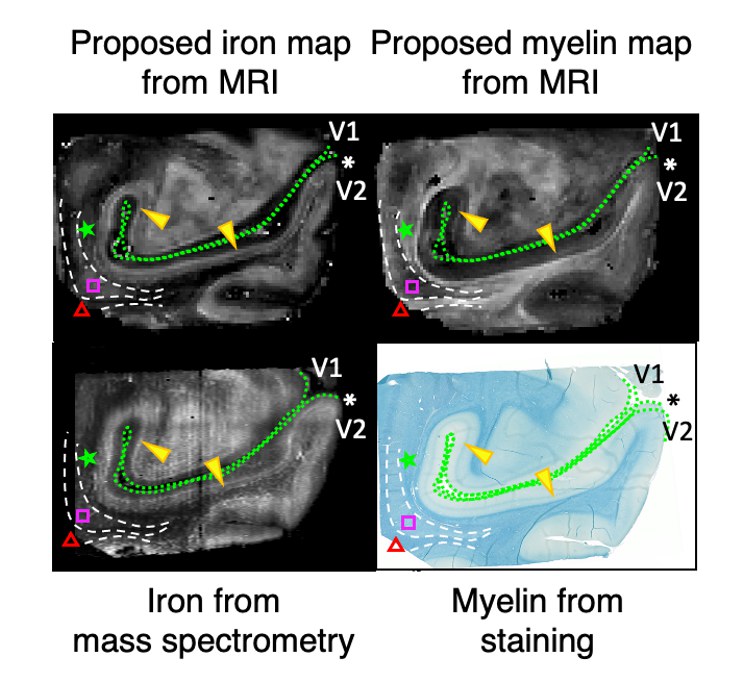
Iron and myelin are two of the main sources of magnetic susceptibility in brain tissue, and both change in many neurological diseases. Iron is paramagnetic and myelin is diamagnetic, so their effects on the MRI signal partly cancel. Conventional quantitative susceptibility mapping (QSM) measures only their sum, which makes a loss of myelin hard to tell apart from a gain in iron.
χ-separation uses a biophysical model of both the frequency shift and the transverse relaxation of the MRI signal to estimate paramagnetic and diamagnetic susceptibility in each voxel. We tested it with simulations and phantoms, compared it with histology in brain specimens, extended it to 7T and to a normative brain atlas, and released it as an open-source toolbox.
Related papers
-
χ-separation: magnetic susceptibility source separation toward iron and myelin mapping in the brain
NeuroImage 240:118371 (2021)
-
A human brain atlas of χ-separation for normative iron and myelin distributions
NMR in Biomedicine 37(12):e5226 (2024)
-
NMR in Biomedicine 37(9):e5167 (2024) *Corresponding authors
Microstructure MRI
An MRI voxel is about a millimeter across and contains several tissue compartments. Water inside axons, between myelin layers and in the extracellular space produces signals with different relaxation and diffusion properties, and separating these signals gives information about structure far below the image resolution.
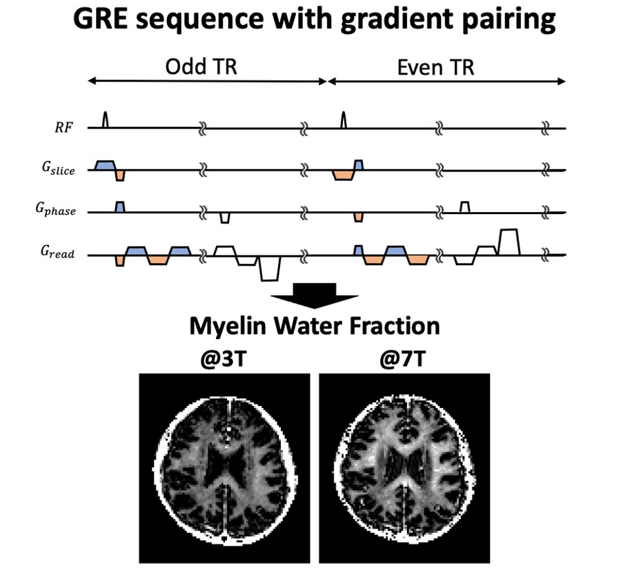
In this area I developed a gradient-echo myelin water imaging method that gives reliable myelin water fraction maps at 3T and 7T. With filtered exchange imaging (FEXI), I showed that the measured water exchange rate in white matter depends strongly on fiber orientation, so FEXI models need to account for compartmental anisotropy. I am now combining susceptibility and myelin water models into a single multi-compartment framework.
Related papers
-
Magnetic Resonance in Medicine 92(2):660-675 (2024)
-
So you want to image myelin using MRI: an overview and practical guide for myelin water imaging
Journal of Magnetic Resonance Imaging 53(2):360-373 (2021)
-
An R2* model of white matter for fiber orientation and myelin concentration
NeuroImage 162:269-275 (2017)
Clinical applications
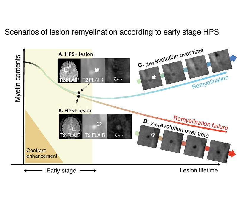
I apply these methods in patient studies with clinical colleagues. In multiple sclerosis, we followed 50 patients for about three years and found that lesions with early iron deposition lost myelin signal over time, while lesions without it recovered. In Parkinson’s disease, χ-separation detected higher iron in the substantia nigra and premotor cortex, a change that conventional susceptibility measures missed.
Other projects include distinguishing multiple sclerosis from neuromyelitis optica spectrum disorder, white matter damage in cerebral small vessel disease, iron accumulation in adrenoleukodystrophy, and brain imaging in post-treatment Lyme disease.
Related papers
-
npj Parkinson's Disease 12(1):200 (2026)
-
NeuroImage: Clinical 45:103748 (2025)
-
Quantifying remyelination using χ-separation in white matter and cortical multiple sclerosis lesions
Neurology 103(6):e209604 (2024)
-
Radiology 307(1):e220941 (2023)
-
Evidence of iron accumulation in cerebral adrenoleukodystrophy: a potential novel disease mechanism
Annals of Clinical and Translational Neurology 13(6):1276-1282 (2026)
Open-source software
-
GitHub
χ-separation toolbox
MATLAB toolbox for magnetic susceptibility source separation, including χ-sepnet. Co-maintained with the Laboratory for Imaging Science and Technology at Seoul National University.