Hyeong-Geol Shin, Ph.D.
신형걸
Assistant Professor
- Department of Neurology, Johns Hopkins University
- F.M. Kirby Research Center for Functional Brain Imaging, Kennedy Krieger Institute
- Microstructure imaging
- Quantitative MRI
- Susceptibility imaging
- Diffusion MRI
- In-vivo histology
- Physics-informed AI
- Google Scholar
- PubMed
- ORCID
- sin4109 (at) gmail (dot) com
About
I am an Assistant Professor in the Department of Neurology at Johns Hopkins University and the F.M. Kirby Research Center for Functional Brain Imaging at Kennedy Krieger Institute.
My research focuses on brain MRI techniques that measure tissue properties related to neurological diseases, including multiple sclerosis, Parkinson’s disease and dementia. I work in three areas. The first is biophysical modeling, which describes how cellular structures shape the MRI signal through magnetic susceptibility, diffusion and water exchange. These models make it possible to measure properties such as myelin content and iron concentration, which conventional MRI cannot quantify. The second is physics-informed artificial intelligence, which builds physical laws into neural networks to reduce errors and make these measurements fast enough for clinical use. The third is clinical translation, which brings these techniques from the laboratory to the bedside through patient studies with clinical colleagues.
Research
More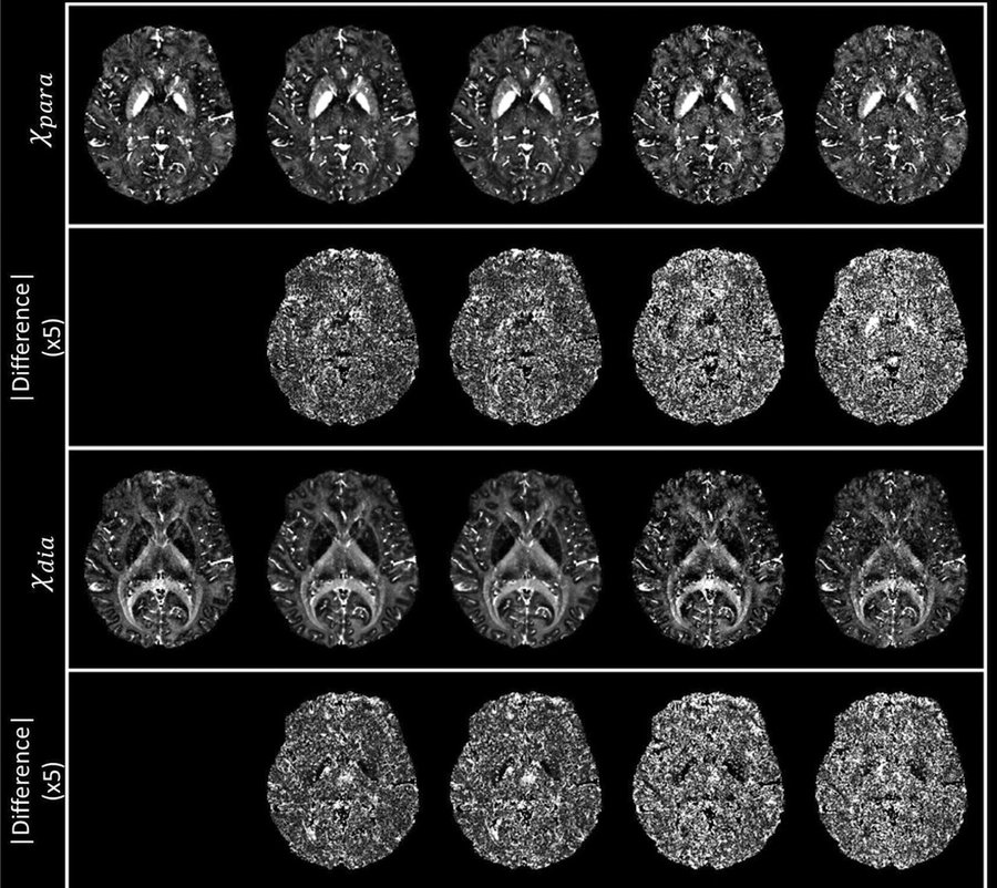
Physics-informed deep learning
Neural networks built on MRI physics for fast and reliable quantitative maps.
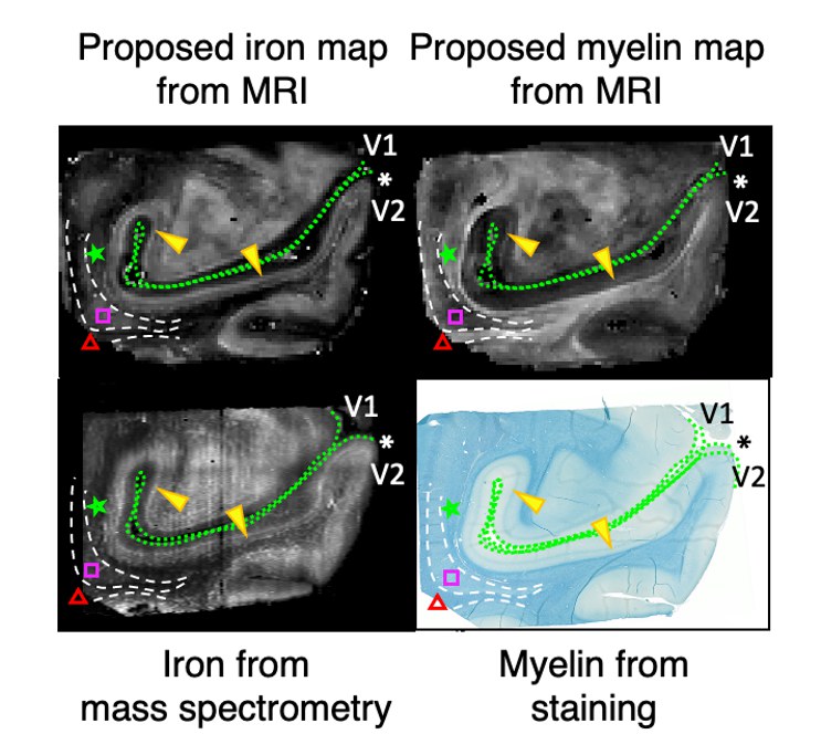
Iron and myelin imaging
Separate maps of iron and myelin, based on a biophysical model of magnetic susceptibility.
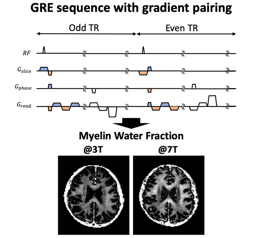
Microstructure MRI
Signals from water in different tissue compartments, used to probe structure below the image resolution.
Clinical applications
Iron and myelin changes in multiple sclerosis, Parkinson's disease and other neurological disorders.
Recent updates
All updates-
Joined the Department of Neurology at Johns Hopkins University as an Assistant Professor. Biography
-
Hosted the 3rd Joint Workshop on MR Phase, Magnetic Susceptibility and Electrical Properties Mapping in Annapolis as a member of the local organizing committee. Service
-
Invited talk, “Update on QSM source separation,” at the 3rd Joint Workshop on MR Phase, Magnetic Susceptibility and Electrical Properties Mapping in Annapolis. Talks
-
Received a National Multiple Sclerosis Society research grant (Co-PI) to study imaging markers of remyelination in multiple sclerosis. Funding
-
Invited talk, “Beyond QSM: separating magnetic susceptibility sources,” at the Gordon Research Conference on In Vivo Magnetic Resonance. Talks
-
Magna Cum Laude Award, ISMRM Annual Meeting, Cape Town. Honors
-
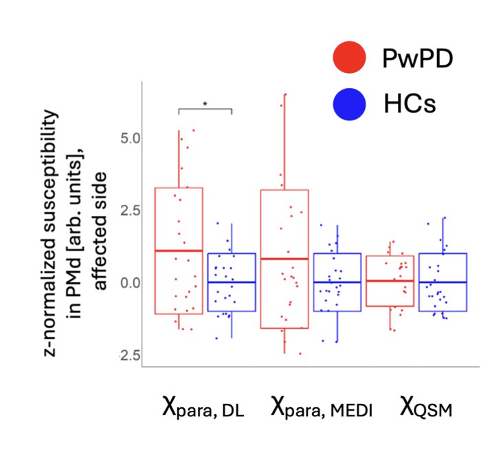
Our study of brain iron in Parkinson’s disease was published in npj Parkinson’s Disease. Paper
-
US Patent 12,411,196 granted, “Method for reconstructing QSM map using dipole compensation.” Patent
-
1st Place Oral Presentation Award, ISMRM White Matter Study Group. Honors
-
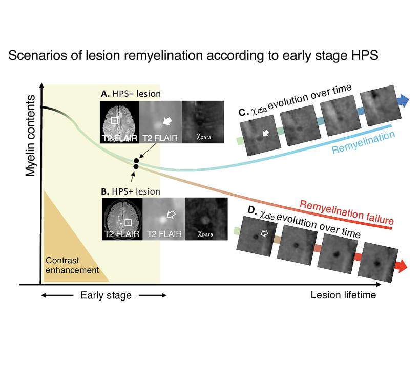
Our study of iron and myelin repair in multiple sclerosis lesions was published in NeuroImage: Clinical. Paper
Selected publications
Full list-
χ-separation: magnetic susceptibility source separation toward iron and myelin mapping in the brain
NeuroImage 240:118371 (2021)
A biophysical model and method that map paramagnetic (iron) and diamagnetic (myelin) susceptibility separately in each voxel.
-
npj Parkinson's Disease 12(1):200 (2026)
In 25 patients and 26 controls, χ-separation found higher iron in the substantia nigra and premotor cortex, which conventional susceptibility maps did not detect.
-
NeuroImage: Clinical 45:103748 (2025)
Over about three years, multiple sclerosis lesions with early iron deposition lost myelin signal, while lesions without it recovered.
-
χ-sepnet: deep neural network for magnetic susceptibility source separation
Human Brain Mapping 46(2):e70136 (2025) *Co-corresponding authors
A neural network that produces χ-separation maps with fewer artifacts, including a version that needs only multi-echo gradient-echo data.
-
Advances in gradient echo myelin water imaging at 3T and 7T
NeuroImage 188:835-844 (2019)
A sequence and processing pipeline for myelin water imaging at 3T and 7T, enabling 1.5 mm isotropic myelin water maps at 7T.